This activity for Diabetic Retinopathy education is provided by Med Learning Group.
This activity is supported by an independent medical education grant from Regeneron Pharmaceuticals, Inc.
Copyright © 2019Med Learning Group. Built by Divigner. All Rights Reserved.
Classifying Diabetic Retinopathy
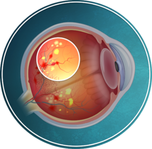
Diabetic retinopathy (DR) is a common complication of diabetes (type 1 or 2).1 It is the manifestation of end-organ damage from diabetes in the eye.2 Classically, DR was considered a microvascular disease of the retina, but newer research suggests neurodegeneration is present before the microvascular anomalies can be seen.1
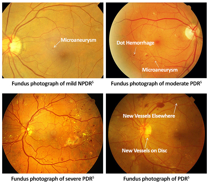
There are 4 stages of diabetic retinopathy3
- Mild nonproliferative diabetic retinopathy (mild NPDR): Small outpouchings of the retinal vascular wall, called microaneurysms, are present in the retina.
- Moderate nonproliferative diabetic retinopathy (moderate NPDR): More microaneurysms are present. Mild vascular changes that affect the shape and structure of the retinal blood vessels, small areas of bleeding (called dot and blot hemorrhages) and soft exudates may also be present in the retina.
- Severe nonproliferative diabetic retinopathy (severe NPDR): Microaneurysms and/or bleeding is present in all areas of the retina and/or more significant vascular changes are present.
- Proliferative diabetic retinopathy (PDR): The growth of fragile, abnormal retinal blood vessels (neovascularization [NV]) is present in the eye. The new blood vessels can grow in the on the optic disc (NVD), iris (NVI), filtration angle (NVA) or retina (NVE).
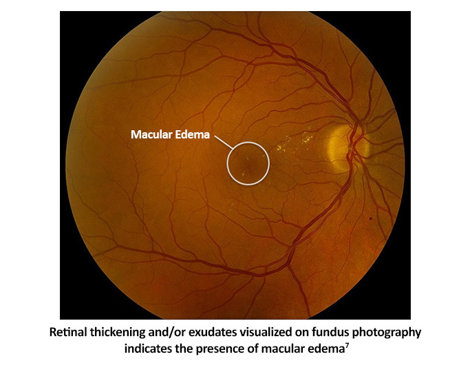
In addition to the stages listed above, the presence or absence of diabetic macular edema (DME) will be documented. DME can develop at any stage of DR. It is swelling in the macula (the area of the retina responsible for central vision) due to leaky, damaged retinal blood vessels.3
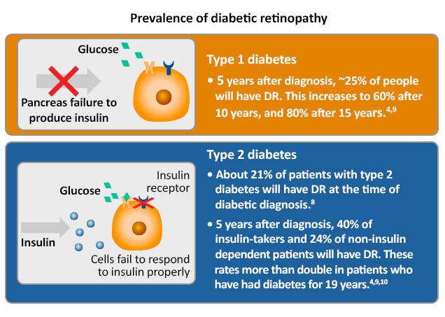
Prevalence of diabetic retinopathy:
References
- Abcouwer SF, Gardner TW. Diabetic retinopathy: Loss of neuroretinal adaptation to the diabetic metabolic environment. Ann N Y Acad Sci. 2014;1377:174-190.
- Shah AR, Garder TW. Diabetic retinopathy: Research to clinical practice. Clin Diabetes Endocrinology. 2017;3:9.
- Grading diabetic retinopathy from stereoscopic color fundus photographs–an extension of the modified Airlie House classification. ETDRS report number 10. Early Treatment Diabetic Retinopathy Study Research Group. Ophthalmology. 1991;98(5 suppl):786-806.
- Flaxel CJ, Adelman RA, Bailey ST, et al. Diabetic Retinopathy Preferred Practice Pattern®. Ophthalmology. 2020;127(1):P66-P145.
- Wong TY, Sun J, Kawasaki R, et al. Guidelines on Diabetic Eye Care: The International Council of Ophthalmology Recommendations for Screening, Follow-up, Referral, and Treatment Based on Resource Settings. Ophthalmology. 2018;125:1608-1622.
- Kaplan HJ. Severe NPDR. Retina Image Bank. 2013;5343. https://imagebank.asrs.org/file/5343/severe-npdr
- Medical Professionals Ophthalmology. Intravitreal anti-VEGF pharmacotherapy safe for treatment of diabetic macular edema. May 2020. https://www.mayoclinic.org/medical-professionals/ophthalmology/news/intravitreal-anti-vegf-pharmacotherapy-safe-for-treatment-of-diabetic-macular-edema/mac-20485933
- Fong DS, Aiello L, Gardner TW, et al. Retinopathy in diabetes. Diabetes Care. 2004;27(supp1):s84-s87.
- Health Style. Diabetes Mellitus. https://www.healthstyle.net.au/article/diabetes-the-tale-of-two-types/
- Klein R, Klein BE, Moss SE, Davis MD, DeMets DL. The Wisconsin Study of Diabetic Retinopathy. II. Prevalence and risk of diabetic retinopathy when age at diagnosis is less than 30 years. Arch Ophthalmology. 1984;102(4):520-526.
All URLs accessed 4/22/22.
